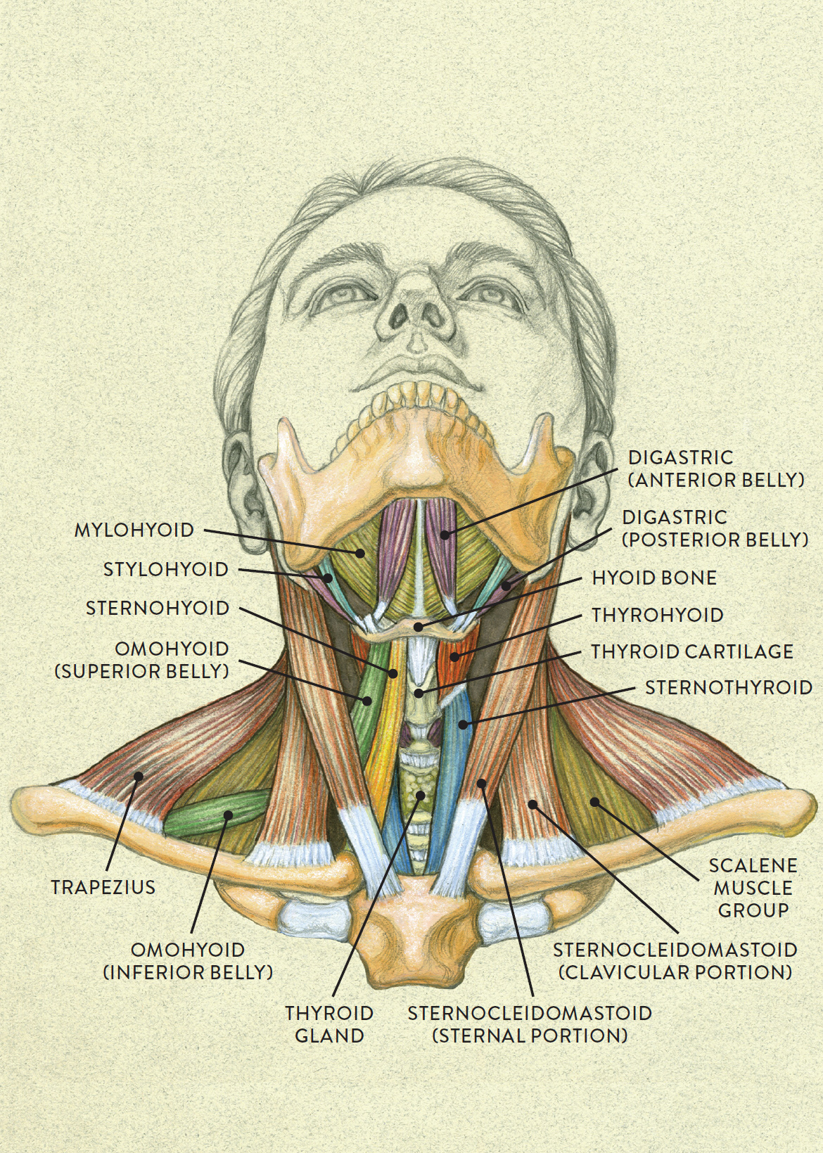
Back Of Neck Anatomy Muscles / / They move the head in every direction, pulling the skull and jaw towards the shoulders, spine, and scapula.. Several other muscles of the back also extend up to the neck region and are partly connected with the cervical part of the vertebral column, including the trapezius, levator scapulae, splenius, iliocostalis, longissimus, rotatores, semispinalis, interspinales, and intertransversarii muscles. Neck muscles help support the cervical spine and contribute to movements of the head, neck, upper back, and posterior longitudinal ligament (pll). Digastric, mylohyoid, geniohyoid, stylohyoid infrahyoid muscles: Many conditions and injuries can affect the back. Sternohyoid, sternothyroid, thyrohyoid, omohyoid anterior vertebral muscles:
The muscles of the anterior neck assist in deglutition (swallowing) and speech by controlling the positions of the larynx (voice box), and the hyoid bone, a the back muscles stabilize and move the vertebral column, and are grouped according to the lengths and direction of the fascicles. It's buried under the sternomastoid anteriorly and by. Posterior and lateral views of the neck by phil schatz. There are several individual muscles within the back anatomy, and it's important to take a quick look the image below to shows all the major back muscles (as well as some neck muscles) Muscles of the shoulder and back laminated anatomy chart.
Anterior view of head tilting back from schoolbag.info Spinous processes of txi to liii and supraspinous ligaments. Cervical spine anatomy is quite complex. The extensors and rotators of the head and neck: Splenius capitis is one of the deep back muscles that is associated with rotating and extending the head and neck. The head rests on the top part of the vertebral column, with the skull joining at c1. The superficial group acts on upper limbs and. Rectus capitis, longus capitis, longus colli. Many in the neck help to stabilize or move the head.
We will attempt to provide a simplified overview of this complex anatomy.
It's buried under the sternomastoid anteriorly and by. The pll starts at c2 and goes down the back of the vertebral bodies and intervertebral discs. Muscles that act on the back. The anterior and middle scalenes originate from the transverse processes of certain cervical vertebrae and attach to the first rib. Superficial muscles are the muscles closest to the skin surface and can usually be seen while a body is performing actions. The posterior muscles of the neck are primarily concerned with head movements, like extension. Splenius capitis is one of the deep back muscles that is associated with rotating and extending the head and neck. Rectus capitis, longus capitis, longus colli. The muscles of the shoulder and back chart shows how the many layers of muscle in the shoulder and back are intertwined with the other relevant systems and muscles in adjacent areas like the spine and neck. Neck muscles help support the cervical spine and contribute to movements of the head, neck, upper back, and posterior longitudinal ligament (pll). 12 photos of the muscle anatomy back of neck. Here the extrinsic back muscles are classified into logical subgroups to facilitate knowledge. Bones of the neck picture.
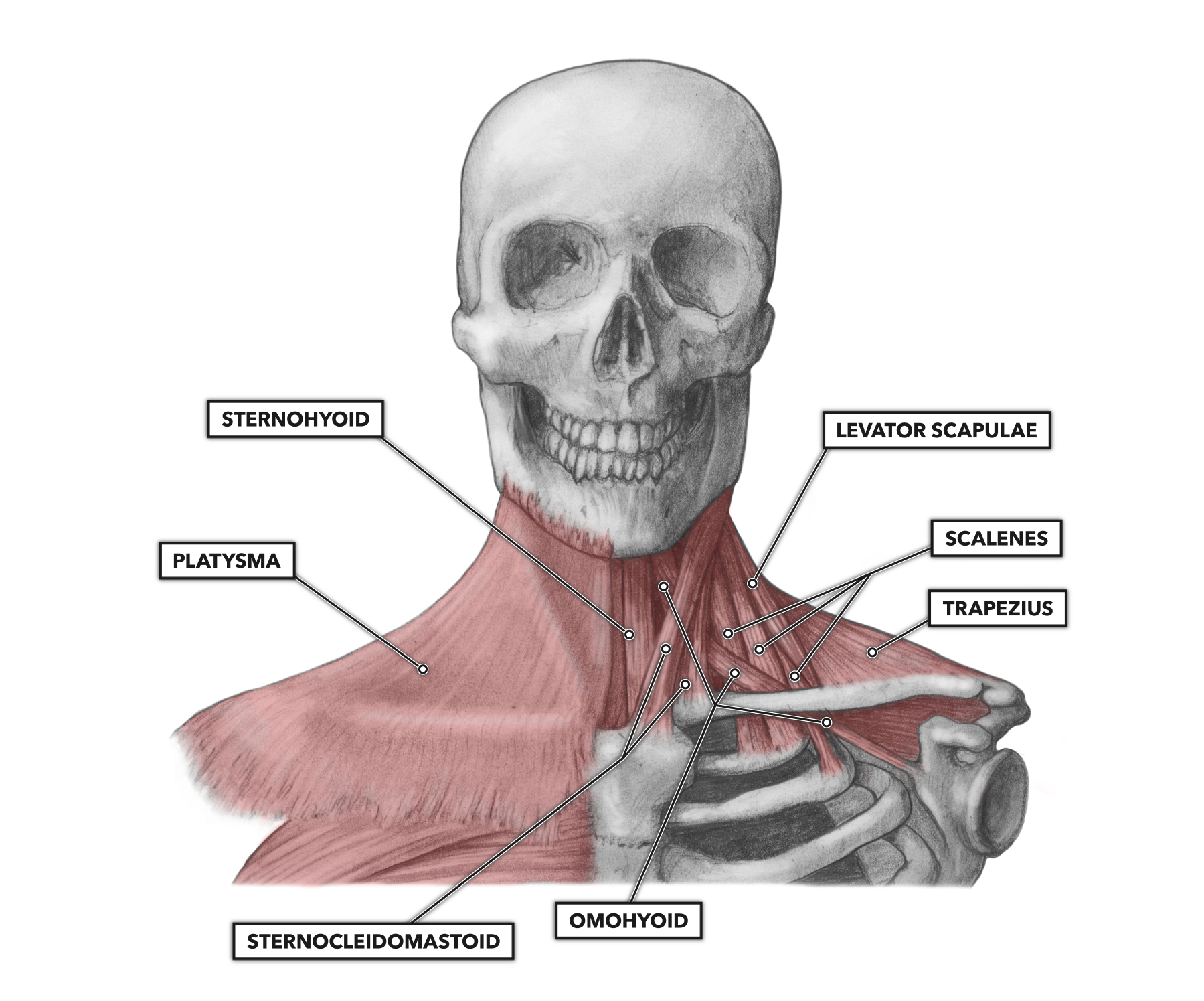
Some neck muscles attach to the clavicles. There are many muscles around the neck that help to support the cervical spine and allow you to move your head in different directions. Many conditions and injuries can affect the back. This article looks at the anatomy of the back, including bones, muscles, and nerves. The three scalene muscles are found forming the floor of the posterior triangle.
Neck muscles from image.slidesharecdn.com The back contains the spinal cord and spinal column, as well as three different muscle groups. The pll starts at c2 and goes down the back of the vertebral bodies and intervertebral discs. It's buried under the sternomastoid anteriorly and by. Only two of the more obvious and superficial neck muscles are. Week 2 anatomy (back/neck muscles). The three scalene muscles are found forming the floor of the posterior triangle. In this section, learn more about the anatomy of the muscles of the neck. This article looks at the anatomy of the back, including bones, muscles, and nerves.
The anterior and middle scalenes originate from the transverse processes of certain cervical vertebrae and attach to the first rib.
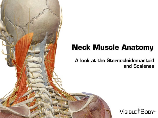
We will attempt to provide a simplified overview of this complex anatomy. The head rests on the top part of the vertebral column, with the skull joining at c1. Digastric, mylohyoid, geniohyoid, stylohyoid infrahyoid muscles: Only two of the more obvious and superficial neck muscles are. The neck muscles (and neck anatomy on the whole) are responsible for head movement, stabilizing the upper region of the body, assisting in the neck muscles include the scalenes, which attach the cervical vertebrae to the thoracic cage, and the sternocleidomastoid, which attaches the skull to the. The major muscle of the back of the neck, the trapezius, is involved in movements of the scapula and is dealt with in the next section, on the muscles in this view of a male figure with one arm up and one arm on the hip, there is a tremendous number of clearly defined anatomical shapes, large and small. Posterior and lateral views of the neck by phil schatz. Intermediate back muscles and c. They start at the top of the neck and go down to the tailbone. The deep back muscles lie immediately adjacent to the vertebral column and ribs. Several other muscles of the back also extend up to the neck region and are partly connected with the cervical part of the vertebral column, including the trapezius, levator scapulae, splenius, iliocostalis, longissimus, rotatores, semispinalis, interspinales, and intertransversarii muscles. The pll starts at c2 and goes down the back of the vertebral bodies and intervertebral discs. This article looks at the anatomy of the back, including bones, muscles, and nerves.
In this section, learn more about the anatomy of the muscles of the neck. Only two of the more obvious and superficial neck muscles are. There are several individual muscles within the back anatomy, and it's important to take a quick look the image below to shows all the major back muscles (as well as some neck muscles) This article gives an overview of the back's structure and its major muscles. The three scalene muscles are found forming the floor of the posterior triangle.
CrossFit | Cervical Muscles, Part 1 from www.crossfit.com The anterior and middle scalenes originate from the transverse processes of certain cervical vertebrae and attach to the first rib. The splenius capitis and cervicis (spinotransversales muscles). They move the head in every direction, pulling the skull and jaw towards the shoulders, spine, and scapula. This article looks at the anatomy of the back, including bones, muscles, and nerves. Sternohyoid, sternothyroid, thyrohyoid, omohyoid anterior vertebral muscles: Lying medial to the iliocostalis muscle group, longissimus is also formed of distinct muscular. Intermediate layer of back muscles. Cervical spine anatomy is quite complex.
The most widely recognized wellsprings of neck torment are.
Remember that there's a small gap between the clavicles where the manubrium sits, about one eyeball if you're having trouble identifying neck muscles, the levator scapulae is the one that points to the ear. Lying medial to the iliocostalis muscle group, longissimus is also formed of distinct muscular. Superficial muscles are the muscles closest to the skin surface and can usually be seen while a body is performing actions. Muscles of the shoulder and back laminated anatomy chart. Muscles that act on the back. Splenius capitis is one of the deep back muscles that is associated with rotating and extending the head and neck. Some neck muscles attach to the clavicles. The back has some of the body's largest muscles (erector spinae group) and some of the smallest what causes neck muscles to tighten, head stuck sideways, eyes opened, jaw locked also back here are the three classes of levers known to physics, with mechanical and anatomical examples. The back muscles stabilize and move the vertebral column, and are grouped according to the lengths and direction of the fascicles. It's buried under the sternomastoid anteriorly and by. This article gives an overview of the back's structure and its major muscles. The muscles of the neck keep running from the base of the skull to the upper back and cooperate to twist the head and help with relaxing. The back anatomy includes the latissimus dorsi, trapezius, erector spinae, rhomboid, and the teres major.
We will attempt to provide a simplified overview of this complex anatomy back of neck anatomy. Splenius capitis is one of the deep back muscles that is associated with rotating and extending the head and neck.


0 Komentar